Reserve your spot for CardioSight.
See how the full experience will work, explore a real sample report, and join the early-access list. No account or password needed — just your name and email.
- Be first in line when analysis opens
- Preview the complete workflow below
- No new scan, no radiation, no commitment
A preview of the full experience
Here's how CardioSight will work at launch.
Every step below is built and waiting. Early-access members unlock the live workflow the moment analysis opens.
01
PreviewPrivacy & consent
See exactly what CardioSight can access, with granular consent you can revoke at any time.
02
PreviewConnect your records
Tell us where your imaging lives — your health system or MyChart portal.
03
PreviewProvide your imaging
Upload an existing chest CT or coronary calcium scan, or authorize us to request it. No new scan, no radiation.
04
PreviewWe build your signal map
Peri-aortic fat is localized and measured, then placed alongside authorized labs and vitals.
05
PreviewYour Heart Age report
Open your result in a secure portal and save a clean, printable PDF to bring to your clinician.
See it for yourself
Explore a sample Heart Age report.
This is exactly the format your own report will take — one clear number, the imaging signal behind it, and next steps for a clinician conversation.
Heart Age
61
yearsReference population
Higher than 82 of 100 reference scans
82nd
Imaging insight
The localized peri-aortic tissue signal resembles an older-appearing cardiovascular phenotype in the research model.
This is not a diagnosis or a prediction of a specific event. It is an imaging-derived reason to place the scan in the context of the person's broader cardiovascular health.
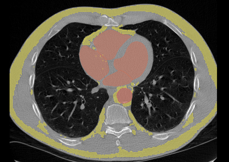
Volume
measuredSpatial burden
Attenuation
measuredTissue density
Texture
measuredHeterogeneity
31 radiomic features -> validated age model -> Heart Age
What can be done next
See where the signal came from, compare it with a reference population, and review common prevention discussion areas with a clinician.
Ready to reserve your spot?
Join the early-access list and we'll reach out the moment your report is ready to run.