Your heart might be older than you are.
See how your heart is aging from a scan you already have — while there is still time to change the story.
CardioSight reads the cardiovascular aging signals hidden in your imaging, labs, and health history, and turns them into one intuitive Heart Age to guide your next conversation with your care team.
Planned early-access price: $99 per report - educational, not a diagnosis
Heart Age
61
yearsChronological age
52
+9 yrs older82nd percentile — a higher cardiovascular aging signal than most of the comparison group.
Imaging signal
82nd
percentile
vs chronological
+9
years older
Model accuracy
±2.7
yrs (MAE)
Educational imaging signal — not a diagnosis. In published research, a higher cardiovascular aging signal has been associated with greater risk.
The unseen problem
You may already have the scan. The information inside it may never have been translated.
A chest CT is usually read for the single reason it was ordered. Your heart, your vessels, and the fat around them hold additional signals about how your cardiovascular system is aging — detail that is not part of a routine read.
That information doesn't disappear. It sits in your imaging, unread and unexplained. CardioSight is built to translate those signals into something you can understand — a single Heart Age.
How it works
From scattered health data to a Heart Age you can use.
CardioSight finds the cardiovascular signals hidden in a scan you already have, puts them in context, and turns the result into a report built for a conversation with your clinician.
01
Your existing scan
Start from a chest CT or heart calcium scan you already have. No new imaging, no radiation.
02
Find your heart & aorta
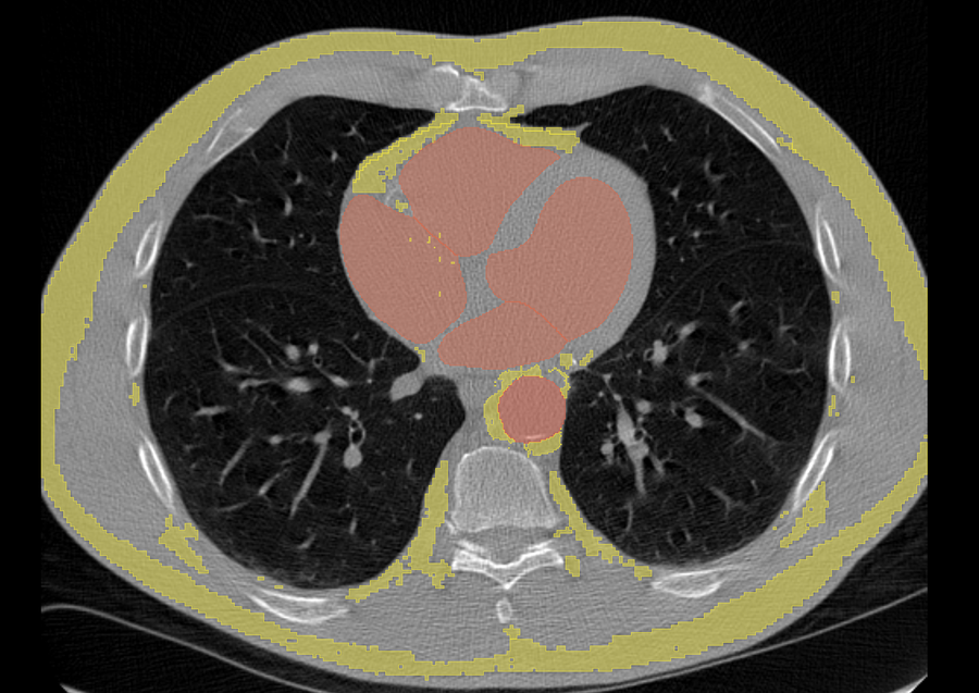
We locate your heart and aorta on the scan so we know exactly where to measure.
03
Measure the fat around them
We measure the fat around the aorta — the proven signal today — and are adding other fat areas over time.
04
Read the tissue
We measure how much fat there is, how inflamed it looks, and its texture.
05
Your Heart Age
Those measurements become one easy-to-understand Heart Age, compared with a reference group.
06
Your report
Your result, the images behind it, and clear next steps arrive in a secure portal and printable PDF.
Heart Age
61
yearsReference population
Higher than 82 of 100 reference scans
82nd
Imaging insight
The localized peri-aortic tissue signal resembles an older-appearing cardiovascular phenotype in the research model.
This is not a diagnosis or a prediction of a specific event. It is an imaging-derived reason to place the scan in the context of the person's broader cardiovascular health.
Volume
measuredSpatial burden
Attenuation
measuredTissue density
Texture
measuredHeterogeneity
31 radiomic features -> validated age model -> Heart Age
What can be done next
See where the signal came from, compare it with a reference population, and review common prevention discussion areas with a clinician.
What you receive
One clear number, explained.
Your report centers on a single Heart Age, the scan signal behind it, and a plain-language explanation of what it can and cannot mean.
- Heart Age. An estimate of how old your heart and vessels look, based on your imaging.
- What was analyzed. Cardiovascular aging signals across your heart, vessels, and surrounding fat — anchored by a validated measure of the fat around the aorta.
- Plain-language explanation. What the result means, what it doesn't, and what to do next.
- A prompt to talk to a clinician. Designed to support — not replace — clinical evaluation.
CardioSight report · illustrative
ExampleHeart Age
61
yearsImaging insight
The fat around the aorta was measured and translated through the validated Heart Age model.
Next: review the result alongside standard cardiovascular risk factors with a clinician.
Illustrative example. Heart Age is an estimate of how old the cardiovascular system appears based on imaging features — not a diagnosis or a prediction of a specific event.
The science
The science behind your Heart Age.
Your Heart Age is grounded in peer-reviewed research on the fat surrounding the aorta — the part of the model that has been validated. We measure how much of that fat there is, how inflamed it looks, and its texture, and we're carefully adding other fat areas, labs, and vitals over time.
Where we look
We use your heart and aorta as landmarks to find and measure the fat around them on the scan you already have.
What we measure
The amount, density, and texture of that fat become the signal behind your Heart Age. Newer signals are clearly labeled as still in development — never presented as proven before they are.
Not a diagnosis
Heart Age is educational information from your imaging. It is designed to support, not replace, a conversation with your clinician.
Tested at scale
The model was trained on 4,451 scans and tested on 44,214 more, across four independent groups of patients.
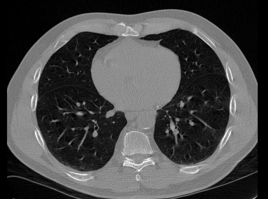
Your existing scan
The chest CT you already have.
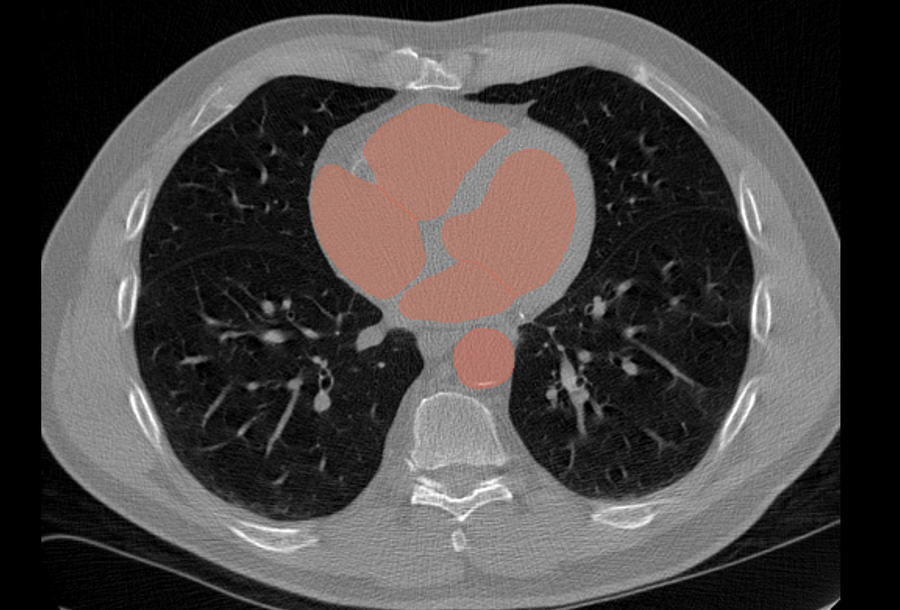
Heart & aorta found
We locate the anatomy to measure around.
Fat measured
The fat around the aorta is isolated and measured.
Primary source
Peri-aortic fat to assess cardiovascular aging using an AI-driven radiomic biomarker
Lebowitz M, Modanwal G, Singh A, et al. European Heart Journal, 2026. A peri-aortic adipose radiomic signature was trained in 4,451 scans and externally validated in 44,214 scans across four CT cohorts, estimating cardiovascular age with a mean absolute error of 2.7 years in the primary external validation.
doi.org/10.1093/eurheartj/ehag498 →Your journey
From curiosity to a clear result.
01
Enter your information
Create a secure account and tell us where your records live.
02
Connect your portal
Choose your health system or MyChart portal so the right record source is identified.
03
Choose how we get the images
Upload the study yourself or authorize CardioSight to request it from the provider.
04
We build your result
Your imaging is measured and placed alongside your labs and vitals.
05
Receive and use the report
Open your Heart Age in the portal, save a PDF, and bring it to a clinician conversation.
Early access uses direct upload or an authorized records request. Direct portal and automated imaging-network retrieval are planned integrations and will only be described as live when they are available.
For health systems
Keep quiet risk from slipping through the cracks.
Give radiology and cardiology teams a way to surface an aging signal from CT studies already moving through care, then route appropriate patients into a follow-up pathway.
Confidence beyond the routine read
Help the care team see a cardiovascular-aging pattern that may not be visible in standard risk fields.
Fewer missed follow-up opportunities
Create a consistent route from an imaging signal to an appropriate clinician review queue.
A workflow clinicians can use
Return Heart Age inside the PACS and health-system workflow rather than in another disconnected portal.
A measurable service line
Track eligible studies, completed analyses, patient outreach, and downstream follow-up without promising a diagnosis.
Why we're building CardioSight
Information that already exists should reach the person it's about.
Millions of people carry a chest CT in their records that contains signal about how their cardiovascular system is aging — signal that is rarely extracted, explained, or connected to prevention. We started CardioSight to close that gap.
CardioSight is led by Mendy Lebowitz, first author of the peri-aortic Cardiovascular Age study published in the European Heart Journal (2026), working with a team of imaging-AI researchers and clinicians.
Privacy by design
Your images stay yours.
We treat your imaging as sensitive health information, we don't sell your data, and you can delete submitted scan and report data from your account.
Encrypted in transit and at rest
Data moves over modern TLS connections and is encrypted at rest by our infrastructure provider.
You control retention
Delete your submitted scan and report data in your account, or contact us to request full account deletion.
Granular, revocable consent
Separate consent for records retrieval, analysis, and any research use — revocable at any time.
No ad trackers on health pages
We don't run advertising pixels on pages that touch your health information.
FAQ
Common questions, straight answers.
- I already had a chest CT — is there cardiovascular information in it I never heard about?
- It may. A chest CT is usually read for the reason it was ordered. CardioSight analyzes imaging patterns across your heart, vessels, and surrounding fat — anchored by a validated peri-aortic measure — that are not part of a routine read.
- Can my cardiovascular system look older or younger than my actual age?
- Yes. Heart Age is an imaging-derived estimate of how old your cardiovascular system appears relative to a research reference population.
- Do I need another scan or more radiation?
- No. CardioSight uses a chest CT or coronary calcium scan you already have — no new imaging and no additional radiation.
- What does the AI analyze?
- It maps your heart, vessels, and the fat around them, then measures imaging features — anchored by a validated measure of the fat around the aorta — that are translated into one Heart Age. More fat areas, labs, and vitals broaden the picture over time.
- Is Heart Age a diagnosis or a prediction that I'll have a heart attack?
- No. It is educational, imaging-derived information — not a diagnosis, not a risk percentage, and not a prediction of a specific event. It's designed to support a conversation with a clinician.
- What if my Heart Age looks accelerated?
- It's a prompt to look closer, not a verdict. Discuss the result with a qualified healthcare professional who can put it in the context of your full health picture.
- Will a clinician review my result?
- An optional clinician consultation is being developed. It is not part of the standard result today, and we don't advertise mandatory clinician review.
- How do you get my scan, and how do I receive the report?
- You upload a scan you have, or we help you request it from your imaging center. Your result is delivered by email and a secure portal. Automated health-record connections are planned, not yet live.
- Who can access my imaging and health information?
- Only the people and systems needed to produce your result. We don't sell your data, we use granular revocable consent, and you can delete submitted scan and report data from your account.
- Is CardioSight FDA-cleared?
- No. CardioSight is not FDA-cleared or approved. It provides educational, imaging-derived information and is not a substitute for professional medical evaluation.
Get a clearer signal while there is still time to act on it.
Join early access, tell us where your records live, and choose whether to upload your CT or authorize us to request it.